AI Microscopy Platform
Control the hardware.
Own the data.
Automate the lab.
A precision adapter, an iPhone, and AI that controls how the image is captured — not just how it's read.
Semen analysis first, then across microscopy.
Clinical partner
CharitéUniversitätsmedizin Berlin
Funded by
Investitionsbank BerlinPro FIT, co-financed by EFRE
Problem
Microscopy diagnostics are
still done by hand.
Most morphology-based diagnostics still depend on a trained human at an eyepiece — which caps quality, capacity and access.
Subjective
The same sample yields different results depending on who reads it. There is no standardization across readers, and none across sites.
Personnel-bound
Every analysis needs a trained reader. Output depends on scarce staff being present, trained and available that day.
Throughput-capped
A reader can sign out only so many analyses per day — often a legal limit as well as a practical one. That is a hard ceiling on capacity.
And today's tools don't close the gap
Unaffordable for most practices
Dedicated analyzers and whole-slide scanners are priced for central labs, not for clinics and practices.
Not performant enough
CASA is weak on morphology. Passive AI scanners have no control over the images they are fed: variable focus, illumination and quality in, unreliable results out.
Approach
We don't just read the image.
We control how it's captured.
A precision adapter, an iPhone and AI on any existing light microscope. Active capture, not passive scanning.
Smartphone
Camera + iOS app
Adapter
Optical coupling
Microscope
Hardware you already own
AI engine
Detection and analysis
Active capture
A camera we control
The iPhone's 48MP computational sensor outresolves the 3.5–5MP cameras most practices use. More importantly, we drive focus, illumination and frame selection: the system decides what gets captured, rather than accepting whatever it is handed. That is the difference between active capture and passive scanning — and it is why the model sees a standardized image every time.
Quality gate
Refuses bad input
Frame quality is scored live, on the device. Poor input is refused rather than analyzed, and the technician is told exactly what to fix.
Data
Data we generate, not scrape
No public or purchased datasets. Our data comes from ethics-approved, SOP-managed clinical studies: standardized capture in, expert-annotated labels out.
Explainable
No black box
Every result is backed by annotated images — marked cells, traceable logic. Reviewers see exactly what the system saw, with a full audit trail.
Adoption
The barrier is a phone clip
A consumer-grade app running on hardware the lab already has. No new instrument to buy, install, learn or find bench space for.
On the bench
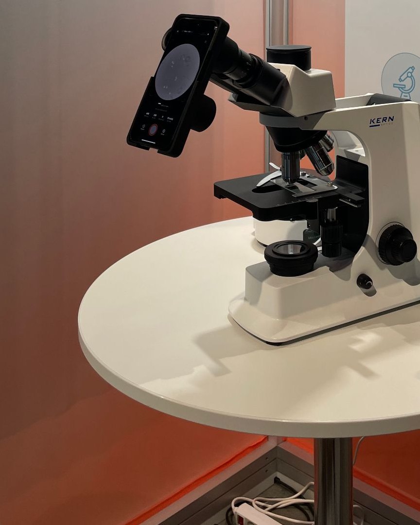
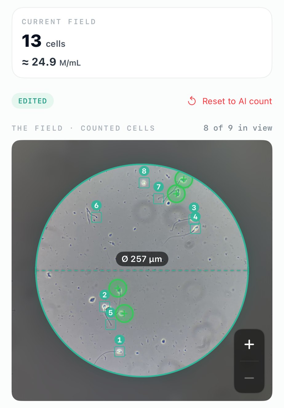
The system, running.
An iPhone on a precision adapter, clipped to a standard light microscope — and the analysis that comes out of it.
First market
Why we start with
semen analysis.
Real, unignorable pain
Manual analysis takes 30–60 minutes per sample, is subjective, and is poorly standardized between readers and sites.
A market CASA validated but left open
CASA proved that clinics will automate this and pay for it. But it fails on morphology, so adoption stayed low. The demand exists; the need is unmet.
The most direct regulatory door
Semen analysis is IVDR Class B — a faster certification path than the Class C modules that follow it.
Per test, against the alternatives
| Metric | Ferto | Manual | CASA |
|---|---|---|---|
| Time per test | ~5 min | 30–60 min | 10–15 min |
| Cost per test | ~€15 | €30–60 | €30–35 |
| Upfront cost | ~€1,000 (adapter + phone) | — | €10,000–30,000 |
| Morphology | Full AI morphology | Slow, subjective | Weak |
CASA cost per test includes roughly €15 of disposables. Ferto figures are planning estimates for the certified product, not measured pilot results.
Platform
A regulated greenfield, on
hardware labs already own.
Where certified tools exist, each one is locked to a dedicated analyzer or a whole-slide scanner in a central lab. None is point-of-care on a standard microscope, and none spans modules.
| Module | Certified today | Bound to | Ferto |
|---|---|---|---|
| Semen analysis | CASA: Hamilton Thorne, Microptic, LensHooke, SQA-V | Dedicated analyzer; weak on morphology | Existing scope, full morphology |
| Urine sediment | Sysmex UF-5000, Beckman iQ200 | Central flow / imaging analyzer | Point-of-care classification |
| Urine cytology | None certified | Manual expert review | Open whitespace |
| Pap / cervical | Hologic Genius, BD, Techcyte | ThinPrep + imager, central lab | No scanner, point-of-care |
| Effusion / FNAC | None certified | Manual expert review | Open whitespace |
| Histology | Paige, Ibex Galen | Whole-slide scanner + digital-path lab | Existing scope |
Semen analysis is the module in development. Every other row is planned, not built — they describe where the platform extends, not what ships today.
Roadmap
The path to Physical Lab AI.
Each certified module is also a data-generation step. The diagnostics business funds the dataset; the dataset trains the model; the model is what eventually runs the hardware.
Phase 1
Passive perception
2026–2028 · Under wayDeploy on tier-one clinical workflows and certify modules in parallel: semen, then urine, then cytology. Quality-gated capture at both ends generates the first structured, gold-standard microscopy dataset.
Output: labelled clinical data
Phase 2
Domain representation
2028–2030 · PlannedOnce several modules reach data density, train a physics-grounded cytology foundation model — one that encodes cellular morphology through optical physics, focal depth and illumination, where general vision models fail.
Output: rich state embeddings
Phase 3
Embodied AI
2030–2033 · PlannedMap those embeddings to motor-control policies. A closed loop drives focus, illumination and stage, so the microscope runs itself — no operator at the eyepiece during analysis.
Output: autonomous analysis
Phase 1 is under way. Phases 2 and 3 are the plan, not built. Each phase is a real business on its own, and the prerequisite for the next.
Regulatory & IP
A dual path, already in motion.
Dual-market from day one
IVDR Class B for semen analysis, and FDA De Novo in parallel. Europe and the US on the same platform, rather than one after the other.
Certify once, then expand
A Predetermined Change Control Plan lets us add modules as controlled changes, without a full re-certification each time.
Reviewed, not theoretical
Our regulatory consultant and the Notified Body have both reviewed and confirmed the approach.
Intellectual property
Core system patent filed
Covering the AI gating mechanism and the inference pipeline. Counsel is actively prosecuting.
Freedom to operate reviewed
No blocking patents identified, with a clear design-around based on our own analysis and system IP.
Pilot programme
Run it in your lab, now.
For fertility clinics, sperm banks and andrology practices. The adapter clips to the microscope already on your bench — no new instrument, no installation, no service contract.
What you get
Adapter, app, support
A precision optical coupler for your existing eyepiece or trinocular port, the capture app, and calibration for your specific scope and objectives.
At the bench
Prepare, clip on, capture
Your existing sample prep, then roughly five minutes per sample. Frame quality is scored live, so if focus or illumination is off you are told what to fix before anything is analyzed.
What you see
Every cell, annotated
Each count comes with annotated images, so you can check what the system found against what you see down the eyepiece.
Interested in a pilot site? Tell us what microscope you run and roughly how many samples a week.
Get in touchWhere we are
Partnered, funded, filed.
Clinical partner
Charité Berlin
Performance study and data collection with Urology and the Kryobank, run under ethics-approved SOPs.
Funding
ProFIT-backed
Public innovation funding from Berlin, awarded to the Ferto–Charité consortium.
Pipeline
RUO pilots
Fertility clinics, sperm banks and andrology practices lined up as research-use pilot sites.
IP
Patent filed
Core system: AI gating and the inference pipeline. Freedom to operate reviewed.
Team
Built health-tech before.
Building the next.

Leo Dieter
CEO & Co-FounderPhD Medical Sciences (Charité), M.Sc. Finance (ESSEC). Five years in digital health, most recently Head of Strategic Projects and employee number one at Kranus Health, where he built the B2B sales motion.
LinkedIn →The rest of the founding team
- Co-Founder & CAIO
- Computer vision and machine learning. Previously an AI data scientist across clinical diagnostics and digital health. Applied mathematics and computer science.
- Co-Founder & CTO
- Mobile systems and regulated medical software. Previously engineering lead at two health-tech companies.
- Medical advisors
- Two senior urologists, between them covering university-hospital practice, clinical trial design and digital health.
Full names and profiles go public shortly. In the meantime — ask us anything.
Contact
Get in touch.
Investors: tell us your focus and we'll send tailored materials on product, roadmap and execution. Clinics and labs: tell us your setup and we'll come back with what a pilot would look like.